[NPE] MEA(Micro Electrode Arrays)
In Vitro Neuron Culture System using MEA
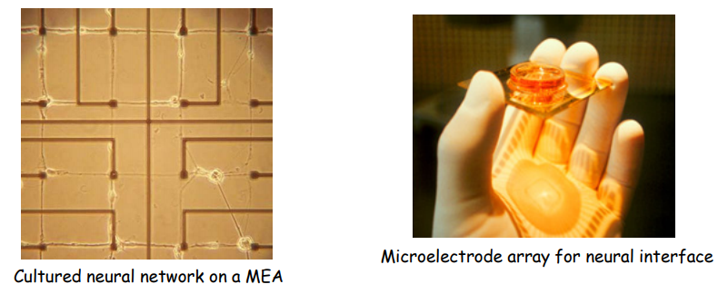
Micro Electrode Arrays
Planar type MEA
- non-invasive
- measuring responses over a longer period of time
- simultaneous multichannel recording
- easy control of the environmental conditions
- easy visualization of cells
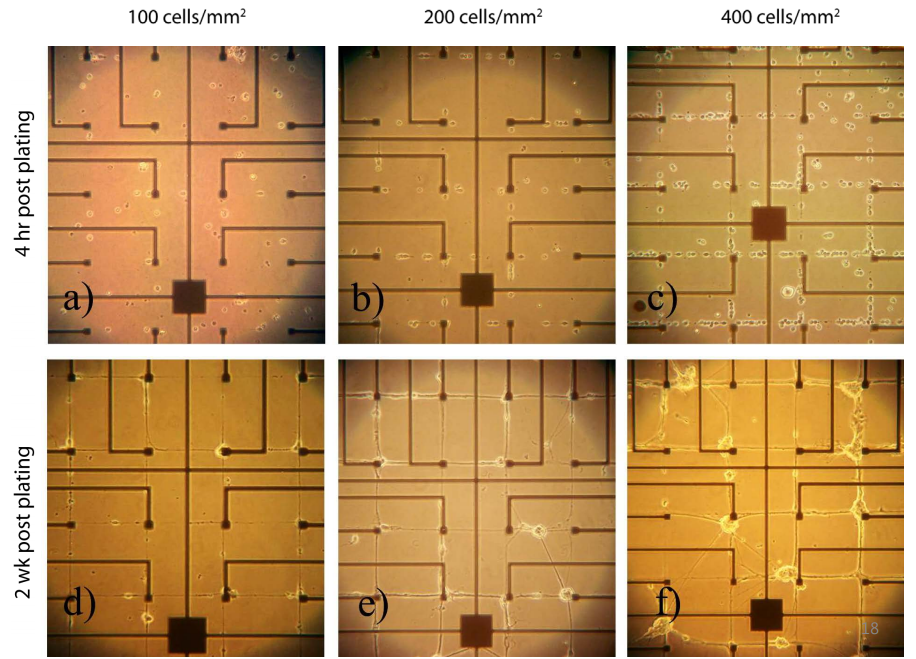
Patterned vs. Random
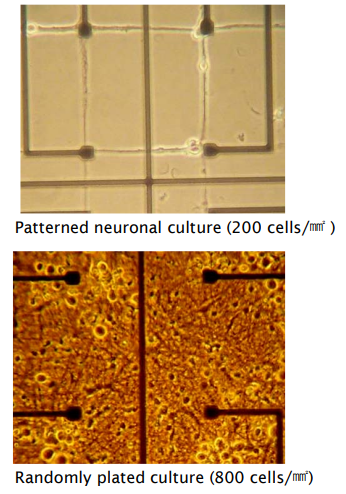
Cellular density in Nueronal network
신경계의 역동학을 정확히 이해하기 위해서는 적절한 밀도의(low density) 잘 짜여진(patterned) 상황을 만들어야 한다.
MEA Fabrication
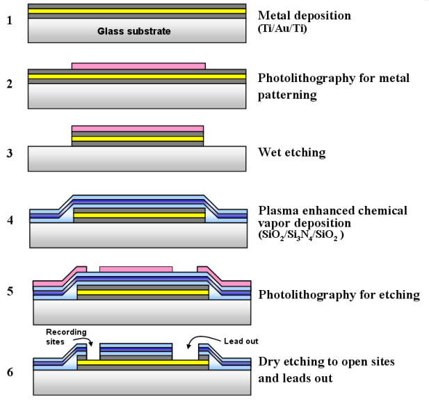
1. Metal deposition
광학현미경 활용을 위해 glass substrate를 쓰고 금을 사용할 경우 접착이 잘 되지 않기 때문에 티타늄을 금 위아래에 추가한다.
2. Phtolithography
우리가 원하는 패턴을 만들기 위해 사진공정을 거친다. 패턴이 그려진 마스크를 올려놓고 강한 빛인 자외선을 쬐면 빛을 받는 부분이 사라진다(= positive photo-resist, 빛은 받지 않는 부분이 사라지기도 한다).
3. Wet etching
4. Insulation layer
유리(SiO2)를 사용하되 Stress-compensation을 위해 Si3N4를 유리 사이에 끼워 넣는다.
5. Etching
6. Dry etching
Insulating layer를 뚫고 전극 site를 위한 금속 layer를 드러나게 한다.
In vitro Neuron Cultures: Cues
Neuron을 원하는 방향으로 자라날 수 있게 Cue를 사용할 수 있다. Protein을 주성분으로 하는 Chemical cues, 기울기 혹은 위치 등을 이용하는 Topographical cues 등이 있다.
Micro-contact Printing: Neural cell adhesion
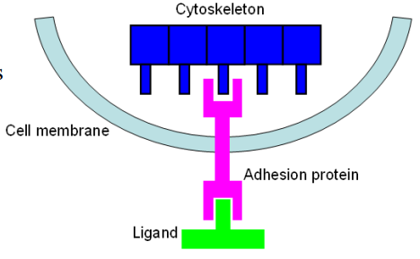
Nueral cell adhesion
Adhesion protein은 ECM 혹은 다른 cell이 발현하는 ligand에 붙으려는 성질이 있다. 이는gene expression, development and growth, survival에 관여한다.
Poly-lysine
- positively charged
- commonly used for non-specific cell binding
- cell attachment by eletrostatic interaction
- protein attachment for cell specific binding
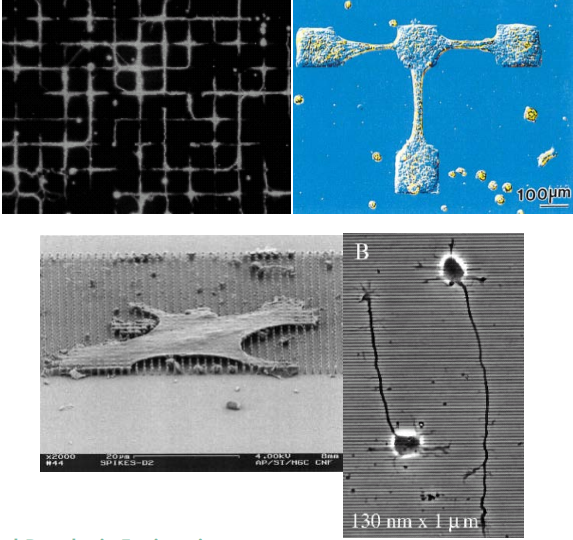
Geometric control of cell attachment
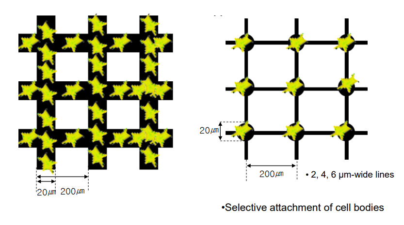
cell body가 cross-section point에만 attachment 되기를 의도하여 디자인 할 수 있다.
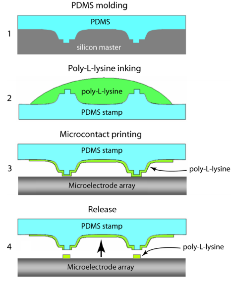
Stamp design : Relief structure
격자 모양으로 패턴화된 MEA 위에 poly-lysine이 coating된 stamp를 격자무늬에 맞춰서 찍는다.
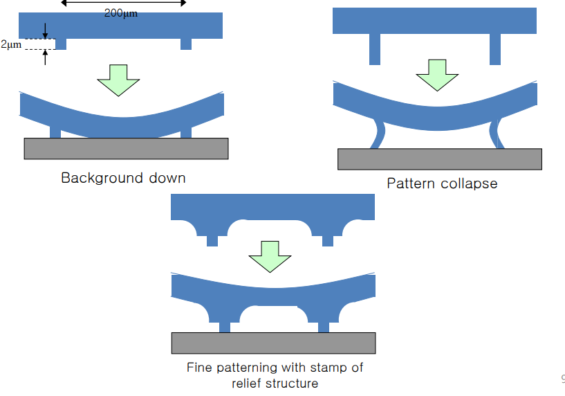
힘을 받는 부위를 relief하는 구조를 만들어 원하는 부위에만 poly-lysine이 묻도록 하는 것이 목표!
Microcontact Priting Procedure(Soft lithography)
Hippocampal Neuron Culture
E18(Embryonic 18 days) hippocampla neurons
18일 경에 가장 많은 viable cell을 얻을 수 있다.
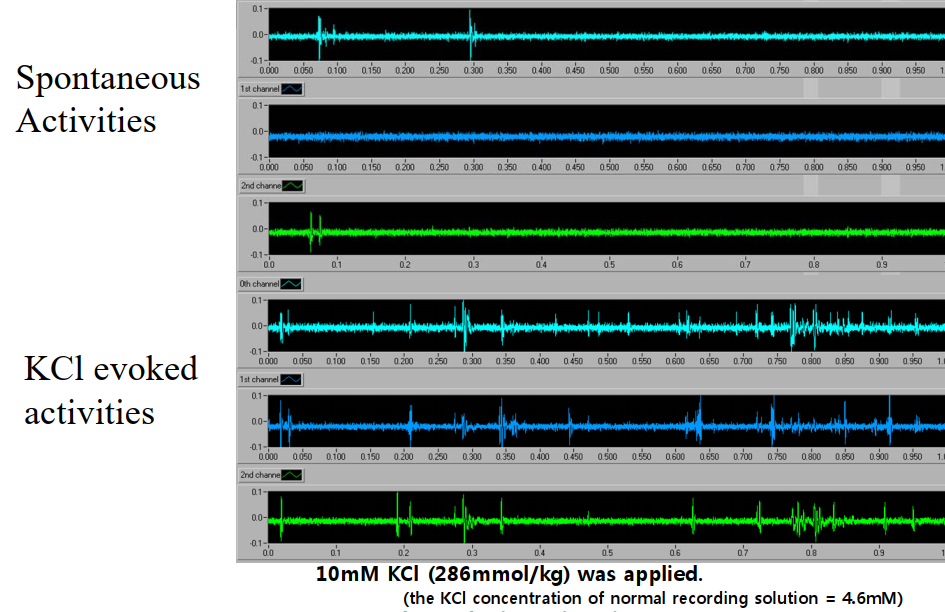
Spontaneous Activity
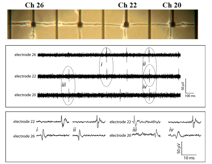
위 사진은 100ms 단위, 아래 사진은 10ms 단위일 때를 보여준다.
i, ii를 살펴보면 항상 26번이 먼저 22번이 나중임을 알 수 있다.
iii, iv를 살펴보면 20번과 22번은 선후관계가 분명치 않음을 알 수 있다.
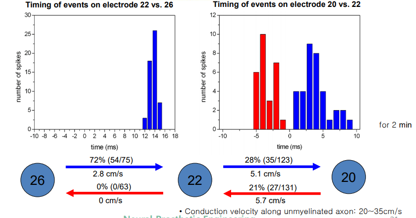
- 살펴보면 배양된 신경세포 cell body 사이의 전파속도가 2~6cm/s 정도로 측정된다. 하지만 실제 단일 unmyelinated axon에서의 전파속도는 20~35cm/s 정도이다. 이를 통해 cell body에서 cell body 사이에 axon 뿐만 아니라 synapse 구조도 존재할 가능성이 있음을 유추할 수 있다.
Electrically Evoked Activity
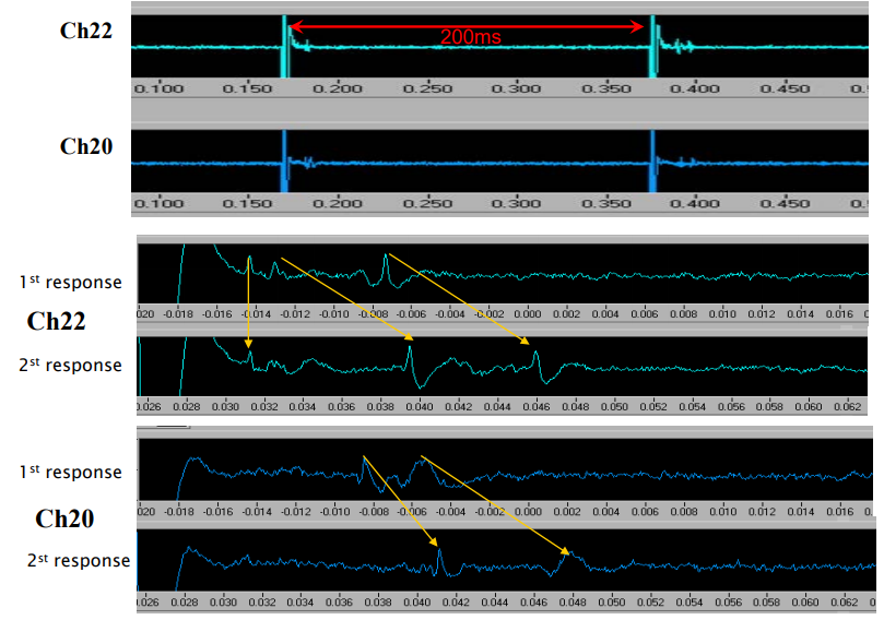
1st stimulation 에서와 달리 2nd stimulation에서는 확실히 response가 느리게 나타난다. 만약 하나의 neuron cell이었다면 (물론 단일 세포의 delayed phase일 가능성도 없진 않지만) 이런 현상이 나타나지 않을 것이다. 이는 neuron cell과 cell 사이인 synapse가 존재할 때 나타는 현상이다. Synapse 사이의 neurotransmitter가 1th stimulation에서 일단 고갈이 되고 다시 충전되는 데에 시간이 걸리기 때문에 2nd stimulation에서 delayed response가 나타나는 것이다.
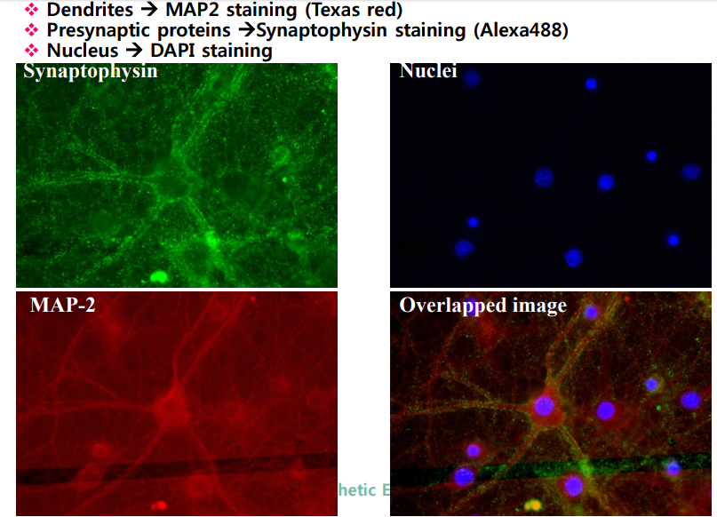
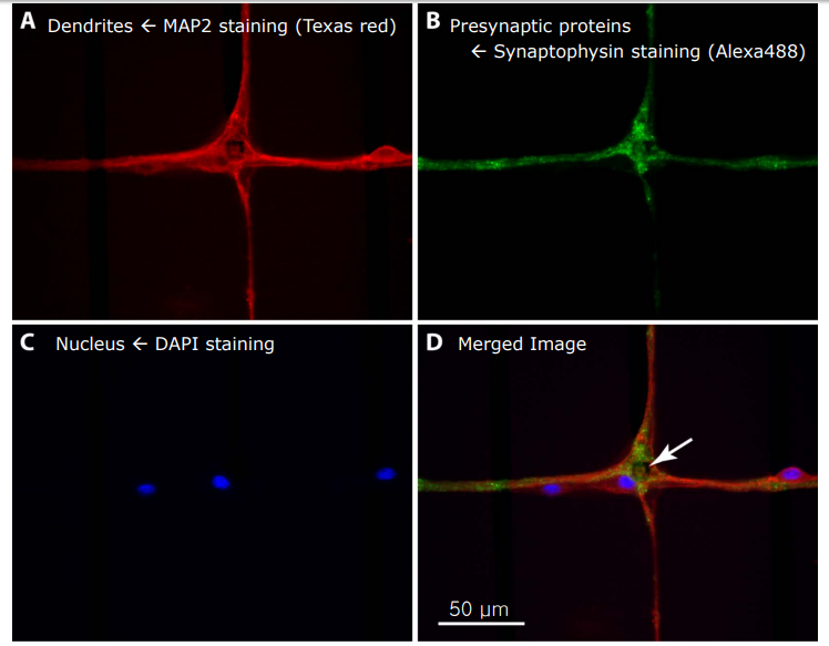
IHC descriptions of cells in networks
In vivo IHC
In patterned network
MEA의 활용
Pharmacology Testing (Toxin Test)
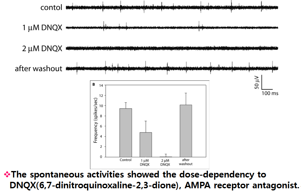
high K+ increases spontaneous activity